Anatomy Of Musckes Sndctendons - Muscle Anatomy & Physiology. Muscle movements, types, and names. Designed for ios, android, windows, and mac. Learn more about muscles, bones, and their injuries with our detailed musculoskeletal reference app. Inflammation of this region caused by repetitive stress or trauma may lead to pain and numbness known as carpal tunnel syndrome. Circular skeletal muscles are made up of fibers explore the minute details of the muscular system in complete anatomy with a suite of 3d learning features such as muscle motion, innervation.
Inflammation of this region caused by repetitive stress or trauma may lead to pain and numbness known as carpal tunnel syndrome. The anterior and middle scalenes originate from the transverse processes of certain cervical vertebrae and attach to the first rib. Learn about human anatomy muscles with free interactive flashcards. In this section, learn more about the anatomy of the muscles of the neck. It occupies most of the oral cavity and oropharynx.
Muscle Anatomy Quiz from www.registerednursern.com An interactive tutorial teaching the position, actions, innervation and attachments of the rectus femoris muscle with the aid of anatomical illustrations. Splenius muscle of head splenius capitis muscle. A detailed guide to understanding how muscles and bones interact, and how common injuries and conditions occur. Skeletal muscles are attached to bones by tendons and can be as long as 30 cm, although they are usually 2 to 3 cm in length. As the skeletal muscles pull on bones to cause movements, they also stabilize the joints of the skeleton; Find the best weight lifting exercises that target each muscle or groups of muscles. The three scalene muscles are found forming the floor of the posterior triangle. The tongue is a mass of muscle that is almost completely covered by a mucous membrane.
By contracting, they also aid the venous return of blood to the heart and with age, these components of the musculoskeletal system progressively degenerate, which contributes to frailty and increases the risk of falls and fractures.
Anatomy of the muscular system. A collection of anatomy notes covering the key anatomy concepts that medical students need to learn. There are around 650 skeletal muscles within the typical human body. Muscular contraction is necessary for voluntary and involuntary movement of limbs, stabilization of joints, maintaining luminal diameter (in the case of arteries, bowel, etc), and to produce heat. Muscle tendons are extremely important in reinforcing and stabilizing joints. The anterior and middle scalenes originate from the transverse processes of certain cervical vertebrae and attach to the first rib. Anatomy of a muscle cell. Inflammation of this region caused by repetitive stress or trauma may lead to pain and numbness known as carpal tunnel syndrome. The three scalene muscles are found forming the floor of the posterior triangle. Anatomy of the short head of the biceps brachii muscle. Muscle mass accounts for a large majority of the carcass weight of domestic animals. How to study muscle anatomy. The tip is the highly mobile, pointed anterior portion of the tongue.
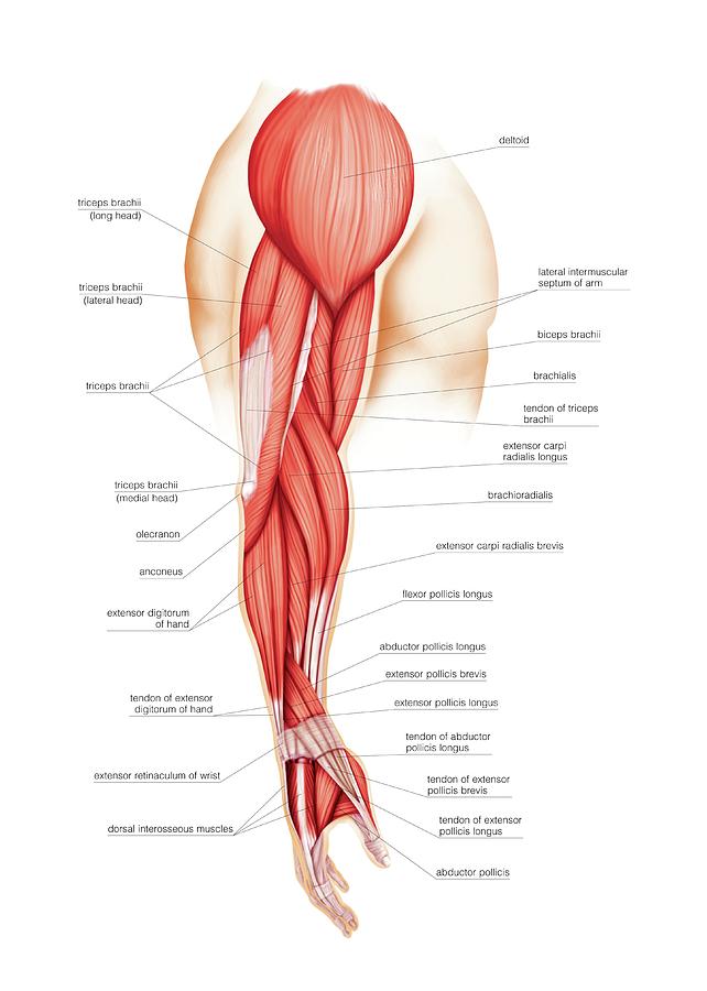
Related online courses on physioplus. The muscles of the torso, examined in the previous chapter, include a few that attach directly into the upper arm and help move the humerus at the shoulder joint. It occupies most of the oral cavity and oropharynx. This article will focus on tongue embryology, origin, insertion, and action of the extrinsic muscles, followed by innervation, blood supply and lymphatic drainage of the tongue. Along with lateral pterygoid muscle it produces side to side movement of mandible.
Back muscles: Anatomy and functions | Kenhub from thumbor.kenhub.com Muscle tendons are extremely important in reinforcing and stabilizing joints. Skeletal muscles allow the body to move and maintain posture; The three scalene muscles are found forming the floor of the posterior triangle. Splenius muscle of head splenius capitis muscle. There are two main muscle groups around the knee: As the skeletal muscles pull on bones to cause movements, they also stabilize the joints of the skeleton; • muscle tissues develop from embryonic cells. Anatomy of a muscle cell.
Skeletal muscles are attached to bones by tendons and can be as long as 30 cm, although they are usually 2 to 3 cm in length.
The anatomy of muscle cells differs from that of other body cells and biologists have applied specific terminology to different parts of these cells. Find the best weight lifting exercises that target each muscle or groups of muscles. The muscular system is responsible for the movement of the human body. An interactive tutorial teaching the position, actions, innervation and attachments of the rectus femoris muscle with the aid of anatomical illustrations. Splenius muscle of head splenius capitis muscle. Lesson on the anatomy of the forearm: Skeletal muscles allow the body to move and maintain posture; It elevates and protrudes the mandible. Learn about human anatomy muscles with free interactive flashcards. This article will focus on tongue embryology, origin, insertion, and action of the extrinsic muscles, followed by innervation, blood supply and lymphatic drainage of the tongue. Muscle tendons are extremely important in reinforcing and stabilizing joints. Muscular contraction is necessary for voluntary and involuntary movement of limbs, stabilization of joints, maintaining luminal diameter (in the case of arteries, bowel, etc), and to produce heat. The tip is the highly mobile, pointed anterior portion of the tongue.
Anatomy of the muscular system. The muscles of the torso, examined in the previous chapter, include a few that attach directly into the upper arm and help move the humerus at the shoulder joint. This article will focus on tongue embryology, origin, insertion, and action of the extrinsic muscles, followed by innervation, blood supply and lymphatic drainage of the tongue. • the muscular system develops from intra embryonic mesoderm. Related online courses on physioplus.
Muscles Of Upper Limb Photograph by Asklepios Medical Atlas from images.fineartamerica.com Muscle mass accounts for a large majority of the carcass weight of domestic animals. Anatomy of a muscle cell. It occupies most of the oral cavity and oropharynx. See the pictures and anatomy description of knee joint bones, cartilage, ligaments, muscle and tendons with resources for knee problems & injuries. There are over two dozen gorgeous and painstakingly. You can click the links in the image, or the links below the image to find out more information on any muscle group. The tongue is a mass of muscle that is almost completely covered by a mucous membrane. Discover the muscle anatomy of every muscle group in the human body.
Discover the muscle anatomy of every muscle group in the human body.
Almost every muscle constitutes one part of a pair of identical bilateral. The tongue is a mass of muscle that is almost completely covered by a mucous membrane. The three scalene muscles are found forming the floor of the posterior triangle. A collection of anatomy notes covering the key anatomy concepts that medical students need to learn. Skeletal muscles allow the body to move and maintain posture; Muscle tendons are extremely important in reinforcing and stabilizing joints. An interactive tutorial teaching the position, actions, innervation and attachments of the rectus femoris muscle with the aid of anatomical illustrations. From anterior to posterior, the tongue has 3 surfaces: Muscles of mastication are classified as main and accessory muscles. The muscular system is responsible for the movement of the human body. This article will focus on tongue embryology, origin, insertion, and action of the extrinsic muscles, followed by innervation, blood supply and lymphatic drainage of the tongue. By contracting, they also aid the venous return of blood to the heart and with age, these components of the musculoskeletal system progressively degenerate, which contributes to frailty and increases the risk of falls and fractures. Microscopic anatomy of skeletal muscle.